Shop
Scoliosis braces


Scoliosis (from Greek: scoliosis meaning from skolios, "crooked" is a medical condition in which a person's spine is curved from side to side. Although it is a complex three-dimensional deformity, on an X-ray, viewed from the rear, the spine of an individual with scoliosis may look more like an "S" or a "C" than a straight line. Scoliosis is typically classified as either congenital (caused by vertebral anomalies present at birth), idiopathic (cause unknown, sub classified as infantile, juvenile, adolescent, or adult, according to when onset occurred), or neuromuscular (having developed as a secondary symptom of another condition, such as spina bifida, cerebral palsy, spinal muscular atrophy, or physical trauma). This condition affects approximately 7 million people in the United States. Patients having reached skeletal maturity are less likely to have a worsening case. Some severe cases of scoliosis can lead to diminishing lung capacity, putting pressure on the heart, and restricting physical activities.
The signs of scoliosis can include:
- Uneven musculature on one side of the spine
- A rib prominence and/or a prominent shoulder blade, caused by rotation of the ribcage in thoracic scoliosis
- Uneven hips/leg lengths
- Slow nerve action (in some cases)
It has been estimated that approximately 65% of scoliosis cases are idiopathic, approximately 15% are congenital and approximately 10% are secondary to a neuromuscular disease. In adolescent idiopathic scoliosis, there is no clear causal agent and it is generally believed to be multifactorial, although genetics are believed to play a role. Various causes have been implicated, but none of them have consensus among scientists as the cause of scoliosis, though the role of genetic factors in the development of this condition is widely accepted. In some cases, scoliosis exists at birth due to a congenital vertebral anomaly. Scoliosis secondary to neuromuscular disease may develop during adolescence, such as with tethered spinal cord syndrome. Scoliosis often presents itself, or worsens, during the adolescence growth spurt and is more often diagnosed in females versus males.

Diagnosis
During the exam, the patient is asked to remove his or her shirt and bend forward. This is known as the Adams Forward Bend and is often performed on school students. If a prominence is noted, then scoliosis is a possibility and the patient should be sent for an X-ray to confirm the diagnosis. It is usual, when scoliosis is suspected, to arrange for weight-bearing full-spine AP/coronal (front-back view) and lateral/sagittal (side view) X-rays to be taken. This is to assess the scoliosis curves and the kyphosis and lordosis, as these can also be affected in individuals with scoliosis. Full-length standing spine X-rays are the standard method for evaluating the severity and progression of the scoliosis, and whether it is congenital or idiopathic in nature. In growing individuals, serial radiographs are obtained at 3–12 month intervals to follow curve progression, and, in some instances, MRI investigation is warranted to look at the spinal cord.
The standard method for assessing the curvature quantitatively is measurement of the Cobb angle, which is the angle between two lines, drawn perpendicular to the upper endplate of the uppermost vertebrae involved and the lower endplate of the lowest vertebrae involved. For patients with two curves, Cobb angles are followed for both curves. In some patients, lateral-bending X-rays are obtained to assess the flexibility of the curves or the primary and compensatory curves.
Management
The traditional medical management of scoliosis is complex and is determined by the severity of the curvature and skeletal maturity, which together help predict the likelihood of progression. The conventional options are, in order:
- Observation
- Physical Therapy
- Bracing
- Surgery
Physiotherapy
The Schroth method is a noninvasive, physiotherapeutic treatment, which has been used successfully in Europe since the 1920s. Originally developed in Germany by scoliosis sufferer Katharina Schroth, this method is now taught to scoliosis patients in clinics specifically devoted to Schroth therapy in Germany, Spain, England, and North America. The method is based upon the concept of scoliosis as resulting from a complex of muscular asymmetries (especially strength imbalances in the back) that can be at least partially corrected by targeted exercises.
Small curvatures between 15 and 20° during growth may be treated with the physio-logic-program. In curvatures exceeding 30°, a combination of the methods described together with the Schroth program may be helpful, and a specialized centre with trained and certified staff should be taken into account. Out-patient rehabilitation treatments today may reach the same outcome as in-patient programs. Out-patient programs may be successful when pattern-specific programs are provided. A certain intensity is necessary to allow the very best compliance with conservative treatment, and to acquire strategies for coping with scoliosis and with the conservative treatment.
The indications for treatment depend on degree of curvature, maturity of the patient, and the individual curve pattern. While evidence supporting such conservative, non-invasive treatments is weak, today conservative management of scoliosis can be regarded as being evidence-based; no substantial evidence has been found to support surgical intervention.

Bracing
Bracing is normally done when the patient has bone growth remaining and is, in general, implemented to hold the curve and prevent it from progressing to the point where surgery is recommended. Braces are sometimes prescribed for adults to relieve pain related to scoliosis. Bracing involves fitting the patient with a device that covers the torso; in some cases, it extends to the neck. The most commonly used brace is a Boston brace (DBB), a corset-like appliance that fits from armpits to hips and is custom-made from fiberglass or plastic. It is sometimes worn 22–23 hours a day, depending on the doctor's prescription, and applies pressure on the curves in the spine. The effectiveness of the brace depends not only on brace design and orthotists skill but on patient compliance and amount of wear per day. The typical use of braces is for idiopathic curves that are not grave enough to warrant surgery, but they may also be used to prevent the progression of more severe curves in young children, to buy the child time to grow before performing surgery, which would prevent further growth in the part of the spine affected.
Bracing may cause emotional and physical discomfort. Physical activity may become more difficult because the brace presses against the stomach, making it difficult to breathe. Children may lose weight from the brace, due to increased pressure on the abdominal area.
The Scoliosis Research Society's recommendations for bracing include curves progressing to larger than 20 degrees, curves presenting between 30 and 45 degrees, Risser Sign 0, 1, or 2 (an X-ray measurement of a pelvic growth area), and less than 6 months from the onset of menses in girls. Progressive scolioses exceeding 25° Cobb in the pubertal growth spurt should be treated with a pattern-specific brace like the Chêneau brace and its derivates with an average brace-wearing time of 16 hours/day (23 hours/day assures the best possible result).
Surgery
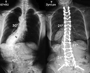
Surgery is usually recommended by orthopedists for curves that have a high likelihood of progression (i.e., greater than 45 to 50 degrees magnitude), curves that would be cosmetically unacceptable as an adult, curves in patients with spina bifida and cerebral palsy that interfere with sitting and care, and curves that affect physiological functions such as breathing.
Surgery for scoliosis is performed by a surgeon specializing in spine surgery. For various reasons, it is usually impossible to completely straighten a scoliotic spine, but in most cases significant corrections are achieved.
There are two main types of surgery:
Anterior fusion: This surgical approach is through an incision (cut) at the side of the chest wall
Posterior fusion: This surgical approach is through an incision on the back and involves the use of metal instrumentation to correct the curve.
One or both of these surgical procedures may be needed. The surgery may be done in one or two stages and, on average, will take four to eight hours.


Our catalog
View our catalog online. 83 product pages for:
- Orthopedics
- Mobility aids
- Splints
- Prosthesis
- Scoliosis
Download it in PDF format